Electron microscopy

Scanning confocal electron microscopy

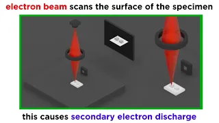
Scanning confocal electron microscopy (SCEM) is an electron microscopy technique analogous to scanning confocal optical microscopy (SCOM). In this technique, the studied sample is illuminated by a focussed electron beam, as in other scanning microscopy techniques, such as scanning transmission electron microscopy or scanning electron microscopy. However, in SCEM, the collection optics is arranged symmetrically to the illumination optics to gather only the electrons that pass the beam focus. This results in superior depth resolution of the imaging. The technique is relatively new and is being actively developed. (Wikipedia).

Scanning confocal electron microscopy
Video thumbnail

DIY Scanning Electron Microscope - Overview

Today, I finally produced an image with my DIY scanning electron microscope. I've spent the last few months working on this project, and am encouraged by today's success. There is still a lot of work left to do in making the image higher resolution, and eliminating sources of noise, howe

From playlist Scanning Electron Microscope

Video thumbnail

DIY Scanning Electron Microscope - Electron Gun Detail

I explain the detailed operation of the electron gun in my DIY scanning electron microscope project.

From playlist Scanning Electron Microscope

Video thumbnail

Electron beam control in a scanning electron microscope

I describe electron beam optics and deflection as they are employed in scanning electron microscopy. http://web.jfet.org/vclk/ - Deflection amplifier http://www.johngineer.com/blog/?p=648 - CRT Christmas tree http://www.fei.com/ - FEI Company

From playlist Scanning Electron Microscope

Video thumbnail

DIY Scanning Electron Microscope - Operation procedure

After getting back from Maker Faire (which is always a hugely enjoyable and inspiring event), I thought that my microscope might need some repairs. As it turned out, I only had to change the filament and tighten some screws that came loose during the trip back from the Faire. The microsc

From playlist Scanning Electron Microscope

Video thumbnail

DIY Scanning Electron Microscope - Sources, Costs and References

Please visit my blog post to see the references and sources for this project: http://benkrasnow.blogspot.com/2011/03/diy-scanning-electron-microscope_26.html

From playlist Scanning Electron Microscope

Video thumbnail

DIY Scanning Electron Microscope - Tektronix contest entry

Vote for me here: http://mytektronixscope.com/videos/ I am using my Tektronix 2246 analog oscilloscope to show the image generated by my DIY electron microscope. The 2246 is operating in X-Y mode, with the channels connected to a raster scan generator. The vertical scanning frequency

From playlist Scanning Electron Microscope

Video thumbnail

Widefield and Confocal Fluorescence Microscopy

We just learned about electron microscopy, so what was the next major innovation in microscopy in the 20th century? That would be fluorescence microscopy, of both the widefield and confocal varieties. How does this work? What is fluorescence in the first place? What are fluorophores? What

From playlist Microbiology/Infectious Diseases

Video thumbnail

Mod-04 Lec-40 Concluding Lecture

Nano structured materials-synthesis, properties, self assembly and applications by Prof. A.K. Ganguli,Department of Nanotechnology,IIT Delhi.For more details on NPTEL visit http://nptel.ac.in

From playlist IIT Delhi: Nano structured materials-synthesis, properties, self assembly and applications | CosmoLearning.org Materials Science

Video thumbnail

Peng Wang - Electron Ptychography: Emerging Computational Microscopy for Physical/Biological Science

Recorded 28 October 2022. Peng Wang of the University of Warwick presents "Electron Ptychography: An Emerging Computational Microscopy for Physical and Biological Sciences" at IPAM's Mathematical Advances for Multi-Dimensional Microscopy Workshop. Abstract: Ptychography is an emerging comp

From playlist 2022 Mathematical Advances for Multi-Dimensional Microscopy

Video thumbnail

Electron Microscopy (TEM and SEM)

We've talked a lot about light microscopy, but this technique has inherent limitations in resolution and magnification. The next paradigm in microscopy that emerged in the middle of the 20th century was electron microscopy. Because electrons have much shorter wavelengths than photons, elec

From playlist Microbiology/Infectious Diseases

Video thumbnail

Microscopes: optical vs SEM vs TEM vs AFM

In order to examine defects and imperfections in materials, we need microscopes capable of enhancing our vision beyond the human eye capability. To access the micro, nano, and atomic scales needed we must rely on different microscopes. The microscope options vary immensely in terms of cost

From playlist Materials Sciences 101 - Introduction to Materials Science & Engineering 2020

Video thumbnail

The Technology of Optical Superoscillations by Nikolay I Zheludev

DISCUSSION MEETING STRUCTURED LIGHT AND SPIN-ORBIT PHOTONICS ORGANIZERS: Bimalendu Deb (IACS Kolkata, India), Tarak Nath Dey (IIT Guwahati, India), Subhasish Dutta Gupta (UOH, TIFR Hyderabad, India) and Nirmalya Ghosh (IISER Kolkata, India) DATE: 29 November 2022 to 02 December 2022 VE

From playlist Structured Light and Spin-Orbit Photonics - Edited

Video thumbnail

Imaging Space Rocks - AMNH SciCafe

What are the technologies at work that help scientists glean information from asteroids and meteors? In this SciCafe, Museum Curator Denton Ebel is joined by Amanda White, a confocal microscopy specialist, and Ellen Crapster-Pregont, a PhD candidate conducting her research at the Museum, i

From playlist SciCafe

Video thumbnail

Pimp your brain: Confocal laser scanning microscopy

In this "pimp your brain" episode Fritz Kragler from the Max Planck Institute of Molecular Plant Physiology in Potsdam-Golm, Germany, talks about fluorescence microscopy, how it works and how it is used in research. In addition, he explains a confocal laser scanning microscope and shows so

From playlist Most popular videos

Video thumbnail

The History of Microscopy

After completing a fairly extensive survey of bacterial and viral pathogens, it's time to learn about some other more general microbiology concepts. What better place to start than the indispensable tool of the microbiologist, the microscope! What is the history of the microscope? What typ

From playlist Microbiology/Infectious Diseases

Video thumbnail

2012 Visualization Challenge: Observing the Coral Symbiome Using Laser Scanning Confocal Microscopy

Christine E. Farrar and colleagues' honorable mention video from the 2012 International Science and Engineering Visualization Challenge, hosted by Science Magazine and the U.S. National Science Foundation, uses confocal microscopy to demonstrate the dynamic lives of corals. [Credit: Chris

From playlist Science & Engineering Visualization Challenge 2012

Video thumbnail

Colin Ophus - Multi-dimensional scanning transmission electron microscopy to solve 3D nanostructures

Recorded 24 October 2022. Colin Ophus of Lawrence Berkeley Laboratory presents "Using multi-dimensional scanning transmission electron microscopy to solve 3D nanostructures using atomic electron tomography" at IPAM's Mathematical Advances for Multi-Dimensional Microscopy Workshop. Abstract

From playlist 2022 Mathematical Advances for Multi-Dimensional Microscopy

Video thumbnail

The Electron Microscope: Where did it come from? | Stuff of Genius

Once we exhausted the ability to magnify objects through the use of light and lenses, we had to look for a new method. Enter the electron microscope. A beam of electrons can get in more of the nooks and crannies of matter to see more detail. Stuff of Genius tells the story behind everyday

From playlist Gizmos & Widgets

Related pages

Scanning electron microscope | Transmission electron microscopy | Scanning transmission electron microscopy | Angular resolution