
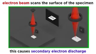
DIY Scanning Electron Microscope - Overview
Today, I finally produced an image with my DIY scanning electron microscope. I've spent the last few months working on this project, and am encouraged by today's success. There is still a lot of work left to do in making the image higher resolution, and eliminating sources of noise, howe
From playlist Scanning Electron Microscope
Electron Microscopy (TEM and SEM)
We've talked a lot about light microscopy, but this technique has inherent limitations in resolution and magnification. The next paradigm in microscopy that emerged in the middle of the 20th century was electron microscopy. Because electrons have much shorter wavelengths than photons, elec
From playlist Microbiology/Infectious Diseases

Chris Russo - Potential electron cryomicroscopy in situ: technology to identify molecules in cells
Recorded 15 November 2022. Chris Russo of the University of Cambridge presents "The potential of electron cryomicroscopy in situ: new technology to identify molecules in cells" at IPAM's Cryo-Electron Microscopy and Beyond Workshop. Abstract: Electron cryomicroscopy (cryoEM) of biological
From playlist 2022 Cryo-Electron Microscopy and Beyond

Mary Scott - Supervised and Unsupervised approaches for Electron Microscopy Data Analysis
Recorded 01 December 2022. Mary Scott of the University of California, Berkeley, presents "Supervised and Unsupervised approaches for Electron Microscopy Data Analysis" at IPAM's Multi-Modal Imaging with Deep Learning and Modeling Workshop. Abstract: Recently, materials science has undergo
From playlist 2022 Multi-Modal Imaging with Deep Learning and Modeling

DIY Scanning Electron Microscope - Electron Gun Detail
I explain the detailed operation of the electron gun in my DIY scanning electron microscope project.
From playlist Scanning Electron Microscope

Anna Gilbert - Imaging from the Inside Out - Inverse Scattering in Fluorescence Microscopy
Recorded 24 October 2022. Anna Gilbert of Yale University presents "Imaging from the Inside Out - Inverse Scattering in Fluorescence Microscopy" at IPAM's Mathematical Advances for Multi-Dimensional Microscopy Workshop. Abstract: We propose a method to reconstruct the optical properties of
From playlist 2022 Mathematical Advances for Multi-Dimensional Microscopy

Peng Wang - Electron Ptychography: Emerging Computational Microscopy for Physical/Biological Science
Recorded 28 October 2022. Peng Wang of the University of Warwick presents "Electron Ptychography: An Emerging Computational Microscopy for Physical and Biological Sciences" at IPAM's Mathematical Advances for Multi-Dimensional Microscopy Workshop. Abstract: Ptychography is an emerging comp
From playlist 2022 Mathematical Advances for Multi-Dimensional Microscopy

Microscope technique brings big resolution at low temperatures
New advances in electron microscopy reveal molecular structures at resolutions useful for drug discovery. Produced by Science and the National Cancer Institute. Animation Credit: Veronica Falconieri and Sriram Subramaniam/LCB/CCR/NCI/NIH Link to article: http://scim.ag/1dYcNi0
From playlist Materials and technology

Microscopes: optical vs SEM vs TEM vs AFM
In order to examine defects and imperfections in materials, we need microscopes capable of enhancing our vision beyond the human eye capability. To access the micro, nano, and atomic scales needed we must rely on different microscopes. The microscope options vary immensely in terms of cost
From playlist Materials Sciences 101 - Introduction to Materials Science & Engineering 2020

Nigel Browning - Inpainting Approaches to Dose Control in High Resolution and In-Situ STEM
Recorded 24 October 2022. Nigel Browning of the University of Liverpool presents "Inpainting Approaches to Dose Control in High Resolution and In-Situ STEM" at IPAM's Mathematical Advances for Multi-Dimensional Microscopy Workshop. Abstract: For many imaging and microanalysis experiments u
From playlist 2022 Mathematical Advances for Multi-Dimensional Microscopy
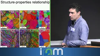
Recorded 25 October 2022. Andy Minor of the University of California, Berkeley, presents at IPAM's Mathematical Advances for Multi-Dimensional Microscopy Workshop. Learn more online at: http://www.ipam.ucla.edu/programs/workshops/workshop-ii-mathematical-advances-for-multi-dimensional-micr
From playlist 2022 Mathematical Advances for Multi-Dimensional Microscopy

Hong Zhou - New cryoEM Programs for Studying Native Biological Complexes, in situ and in Action
Recorded 15 September 2022. Hong Zhou of the University of California, Los Angeles, presents "New cryoEM Programs for Studying Native Biological Complexes, in situ and in Action" at IPAM's Computational Microscopy Tutorials. Abstract: Z. Hong Zhou1,2 1California NanoSystems Institute, Uni
From playlist Tutorials: Computational Microscopy 2022

Investigating the structure of molecules inside cells
International Lecture given by Professor Wolfgang Baumeister. Professor Baumeister will discuss a method, cryo-electrontomography, that has the unique potential to study the ‘molecular sociology’ of cells, combining the best structural preservation with 3D high resolution imaging.
From playlist Latest talks and lectures

Sandra van Aert - 3D atomic resolution through dose-efficient fusion of image & analytical technique
Recorded 26 October 2022. Sandra van Aert of the University of Antwerp presents "3D atomic resolution reconstructions through dose-efficient fusion of imaging techniques and analytical techniques in quantitative STEM" at IPAM's Mathematical Advances for Multi-Dimensional Microscopy Worksho
From playlist 2022 Mathematical Advances for Multi-Dimensional Microscopy

Nikolaus Grigorieff - Detecting 60S Ribosome Maturation Intermediates in Cells by 2D Template Match
Recorded 15 November 2022. Nikolaus Grigorieff of the University of Massachusetts Medical School presents "Detecting Distinct 60S Ribosome Maturation Intermediates in Cells by 2D Template Matching" at IPAM's Cryo-Electron Microscopy and Beyond Workshop. Abstract: For a full understanding o
From playlist 2022 Cryo-Electron Microscopy and Beyond

Fluorescent microscopes are amazing!
Fluorescent microscopy was always one of my favorite parts of working with mammalian cells as it always made for spectacular images. There are a variety of techniques that make use of fluorescence microscopy, many of which are used regularly in the best labs in the world. But fluorescence
From playlist Biology and Genetics
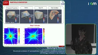
Marie-Ingrid Richard - Structural evolution of nanoparticles observed with Bragg coherent x-ray
Recorded 12 October 2022. Marie-Ingrid Richard of the Commissariat à l'Énergie Atomique (CEA) presents "Structural evolution of nanoparticles under realistic conditions observed with Bragg coherent x-ray imaging" at IPAM's Diffractive Imaging with Phase Retrieval Workshop. Abstract: The ad
From playlist 2022 Diffractive Imaging with Phase Retrieval - - Computational Microscopy