Multidimensional signal processing | Electron microscopy

Electron tomography

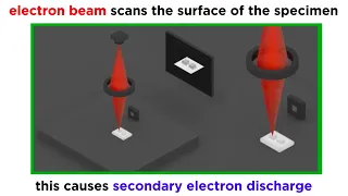
Electron tomography (ET) is a tomography technique for obtaining detailed 3D structures of sub-cellular, macro-molecular, or materials specimens. Electron tomography is an extension of traditional transmission electron microscopy and uses a transmission electron microscope to collect the data. In the process, a beam of electrons is passed through the sample at incremental degrees of rotation around the center of the target sample. This information is collected and used to assemble a three-dimensional image of the target. For biological applications, the typical resolution of ET systems are in the 5–20 nm range, suitable for examining supra-molecular multi-protein structures, although not the secondary and tertiary structure of an individual protein or polypeptide. (Wikipedia).

Electron tomography
Video thumbnail

DIY Scanning Electron Microscope - Overview

Today, I finally produced an image with my DIY scanning electron microscope. I've spent the last few months working on this project, and am encouraged by today's success. There is still a lot of work left to do in making the image higher resolution, and eliminating sources of noise, howe

From playlist Scanning Electron Microscope

Video thumbnail

DIY Scanning Electron Microscope - Electron Gun Detail

I explain the detailed operation of the electron gun in my DIY scanning electron microscope project.

From playlist Scanning Electron Microscope

Video thumbnail

Electron Microscopy (TEM and SEM)

We've talked a lot about light microscopy, but this technique has inherent limitations in resolution and magnification. The next paradigm in microscopy that emerged in the middle of the 20th century was electron microscopy. Because electrons have much shorter wavelengths than photons, elec

From playlist Microbiology/Infectious Diseases

Video thumbnail

ultrasound - A scans explained

Reviews how an A amplitude (A) scan is produced in the context of ultrasound/sonograms See www.physicshigh.com for all my videos and other resources. If you like this video, please press the LIKE and SHARE with your peers. And please add a COMMENT to let me know I have helped you. Follow

From playlist Medical Physics

Video thumbnail

The Holometer: A Fermilab Experiment

Do we live in a two-dimensional hologram? A group of Fermilab scientists has designed an experiment to find out. It’s called the Holometer, and this video gives you a behind-the-scenes look at the device that could change the way we see the universe. Find out more at http://holometer.fnal.

From playlist Detectors and Accelerators

Video thumbnail

Yongsoo Yang - Neural network-assisted atomic electron tomography - IPAM at UCLA

Recorded 26 October 2022. Yongsoo Yang of the Korea Advanced Institute of Science and Technology presents "Neural network-assisted atomic electron tomography" at IPAM's Mathematical Advances for Multi-Dimensional Microscopy Workshop. Abstract: Functional properties of nanomaterials strongl

From playlist 2022 Mathematical Advances for Multi-Dimensional Microscopy

Video thumbnail

Peng Wang - Electron Ptychography: Emerging Computational Microscopy for Physical/Biological Science

Recorded 28 October 2022. Peng Wang of the University of Warwick presents "Electron Ptychography: An Emerging Computational Microscopy for Physical and Biological Sciences" at IPAM's Mathematical Advances for Multi-Dimensional Microscopy Workshop. Abstract: Ptychography is an emerging comp

From playlist 2022 Mathematical Advances for Multi-Dimensional Microscopy

Video thumbnail

Hologram Project!!!

This video is used for Hologram technology, just make the hologram device at home with a very simple way, I'll put a video of how to make the Hologram device. Enjoy!

From playlist OPTICS

Video thumbnail

Manuel Guizar-Sicairos - Resonant ptychography, 3D magnetization and chemical characterization

Recorded 10 October 2022. Manuel Guizar-Sicairos of the Paul Scherrer Institute presents "Resonant ptychography, applications to 3D magnetization and chemical characterization" at IPAM's Diffractive Imaging with Phase Retrieval Workshop. Abstract: Ptychography is an imaging technique that

From playlist 2022 Diffractive Imaging with Phase Retrieval - - Computational Microscopy

Video thumbnail

AWESOME EXPERIMENT Kirlian photography!!!

In this video i show how to take Kirlian photographs with high voltage rumkorff coil. Enjoy!

From playlist ATOMIC PHYSICS

Video thumbnail

Physics 3C. Lecture 26.

UCI Physics 3C: Basic Physics III (Fall 2013) Lec 26. Basic Physics III View the complete course: http://ocw.uci.edu/courses/physics_3c_basic_physics_iii.html Instructor: Michael Smy, Ph.D. License: Creative Commons CC-BY-SA Terms of Use: http://ocw.uci.edu/info More courses at http://ocw

From playlist Physics 3C: Basic Physics III

Video thumbnail

Alberto Bartesaghi - Imaging biomolecules & high-resolution single-particle cryo-electron microscopy

Recorded 13 October 2022. Alberto Bartesaghi of Duke University presents "Imaging biomolecules using high-resolution single-particle cryo-electron microscopy" at IPAM's Diffractive Imaging with Phase Retrieval Workshop. Abstract: Single-particle cryo-electron microscopy (EM) is a popular t

From playlist 2022 Diffractive Imaging with Phase Retrieval - - Computational Microscopy

Video thumbnail

The Electron Microscope: Where did it come from? | Stuff of Genius

Once we exhausted the ability to magnify objects through the use of light and lenses, we had to look for a new method. Enter the electron microscope. A beam of electrons can get in more of the nooks and crannies of matter to see more detail. Stuff of Genius tells the story behind everyday

From playlist Gizmos & Widgets

Video thumbnail

How do PET scans work to detect things such as cancer?

In this video, I discuss Positron Emission Tomography. In particular, I refer to the source of the positron, its annihilation and how the resultant gamma rays are collected. I also briefly discuss its diagnostic benefits. See more Medical imaging techniques here - https://www.youtube.co

From playlist Medical Physics

Video thumbnail

Stimulated Emission Explained

https://www.patreon.com/edmundsj If you want to see more of these videos, or would like to say thanks for this one, the best way you can do that is by becoming a patron - see the link above :). And a huge thank you to all my existing patrons - you make these videos possible. In this video

From playlist Optoelectronic and Photonic Devices

Video thumbnail

Sebastian Seung - Petascale connectomics and beyond - IPAM at UCLA

Recorded 10 October 2022. Sebastian Seung of Princeton University presents "Petascale connectomics and beyond" at IPAM's Diffractive Imaging with Phase Retrieval Workshop. Abstract: A connectome represents brain connectivity as a directed graph in which nodes are neurons and edges are syna

From playlist 2022 Diffractive Imaging with Phase Retrieval - - Computational Microscopy

Related pages

Atomic number | Three-dimensional space | Scanning transmission electron microscopy | Crowther criterion | Electron | Transmission electron microscopy | High-resolution transmission electron microscopy | Tomographic reconstruction